The clinical half of this resource: what each investigation can reasonably answer, the professional standards that govern reporting, the statutory duties attached to dose and data, and how images should be handled.
Radiology library
This section covers what each imaging modality can reasonably answer, the professional standards governing the interpretation and reporting of imaging, and the statutory requirements for dose, quality assurance and image handling.
Providing a clear, practical overview of day-to-day radiological principles for non-specialist readers, trainees, and experienced practitioners seeking a concise reference.Last reviewed September 2026
§ 01 Modalities
Common radiology examinations
Each modality has its own strengths, weaknesses and clinical risks. The following sections describe what each modality can reasonably answer, where it is limited, and the practical safety considerations.
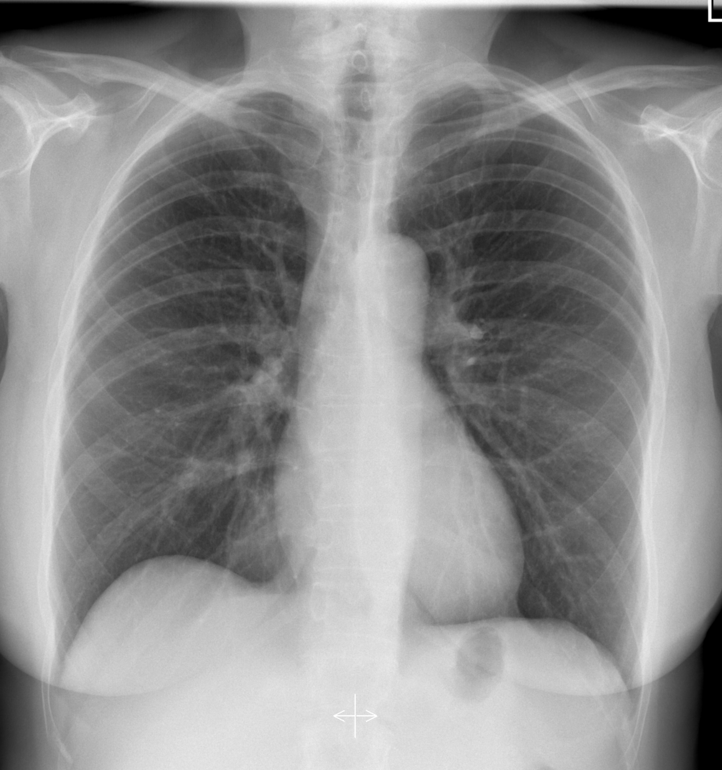
Radiography (X-ray)
Excellent for rapid triage and diagnosis
The classic radiology examination, the plain radiograph, has
historically been the mainstay of most radiology departments, but
in recent years lower-dose CT scanners and increasingly detailed
MRI have gradually begun to supplant this old, reliable workhorse.
However, plain radiographs remain extremely useful and are readily
available in almost every healthcare setting. For some
examinations, a plain radiograph provides all the necessary
information in a single image, allowing a faster and easier
diagnosis that is no less accurate. In some situations, however, a
plain radiograph is much less accurate than CT or MRI. Despite
their relatively low radiation dose, plain radiographs will remain
a vital part of any radiology department for many years to come.
Common indications
Suspected fracture or dislocation
Chest infection, consolidation or pleural effusion
Bowel obstruction or free intraperitoneal gas
Foreign-body localisation
Limitations
Superimposed structures can mask or mimic pathology
Low soft-tissue contrast compared with CT or MRI
Projection geometry can conceal a fracture line
Safety and practical notes
Low-dose examination. Pregnancy status should be checked where
the abdomen or pelvis is in the primary beam. Gonad shielding is
no longer indicated under modern collimation.
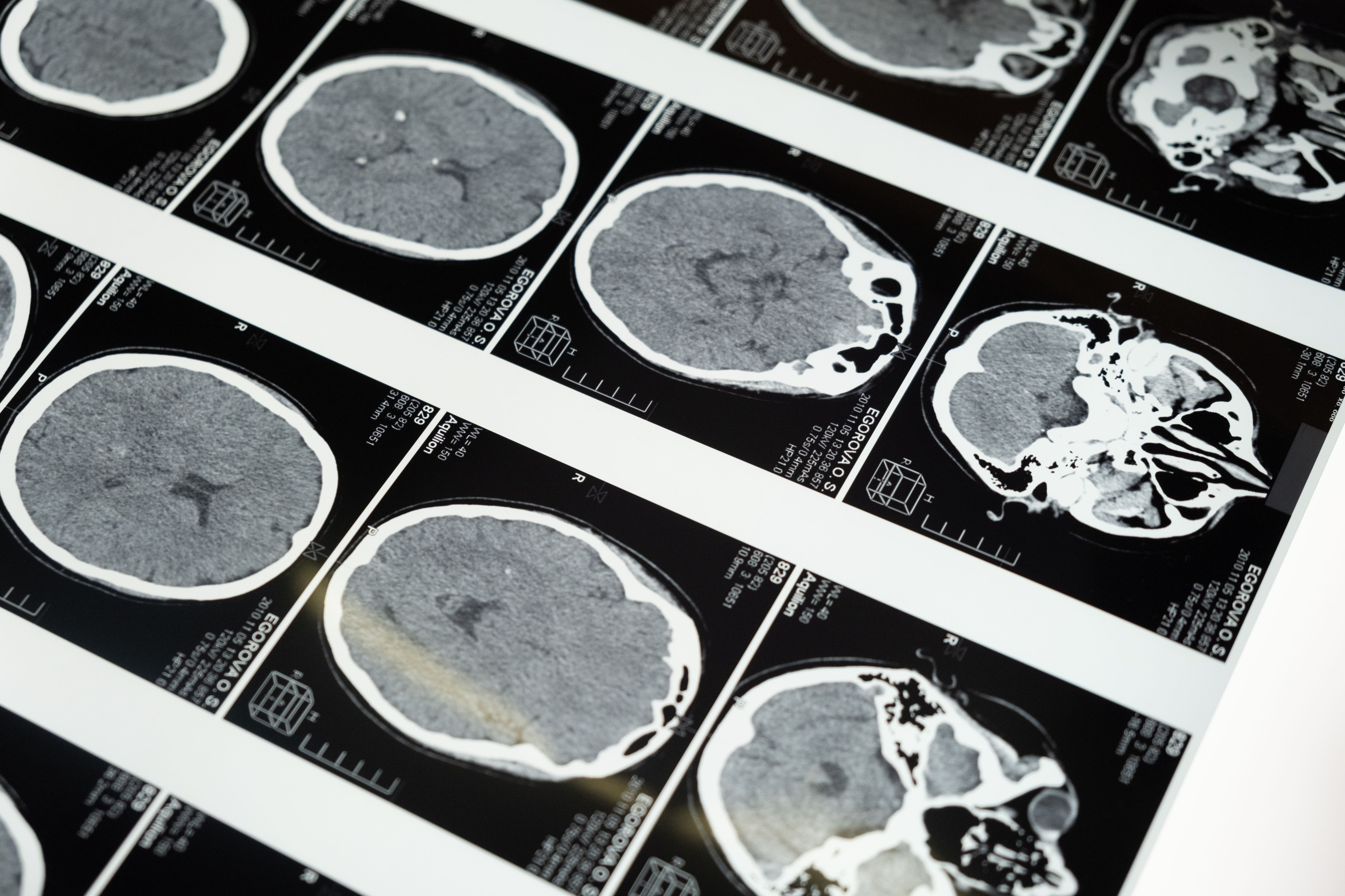
Computed tomography (CT)
Excellent for most acute and cross-sectional questions
Computed tomography is the modern workhorse of acute and
cross-sectional imaging, with high spatial resolution, short
acquisition times and near-universal availability in secondary
care. Multi-detector scanners can image the whole chest and
abdomen in seconds, which is why CT is first line for acute
presentations, trauma, most suspected malignancies and vascular
disease. Its main weakness is that it uses ionising radiation at
doses higher than those of plain radiography, and it often
requires iodinated contrast (an iodine-based agent injected to
make structures easier to see), which carries risks of allergy and
renal injury. Other technical issues, such as beam-hardening and
metal artefact, can also obscure detail -- an issue less prevalent
with plain radiographs. Regardless, every exposure must be
justified under IR(ME)R.
Common indications
Acute head injury, stroke or intracranial haemorrhage
Acute abdomen, trauma and suspected perforation
Pulmonary embolism and aortic dissection
Staging and follow-up of malignancy
Limitations
Ionising radiation dose -- justification and optimisation
apply
Beam-hardening and metal artefact can obscure detail
Safety and practical notes
Justification is mandatory under IR(ME)R. Contrast-enhanced
studies require recent renal function and allergy history. Dose
is monitored against diagnostic reference levels.
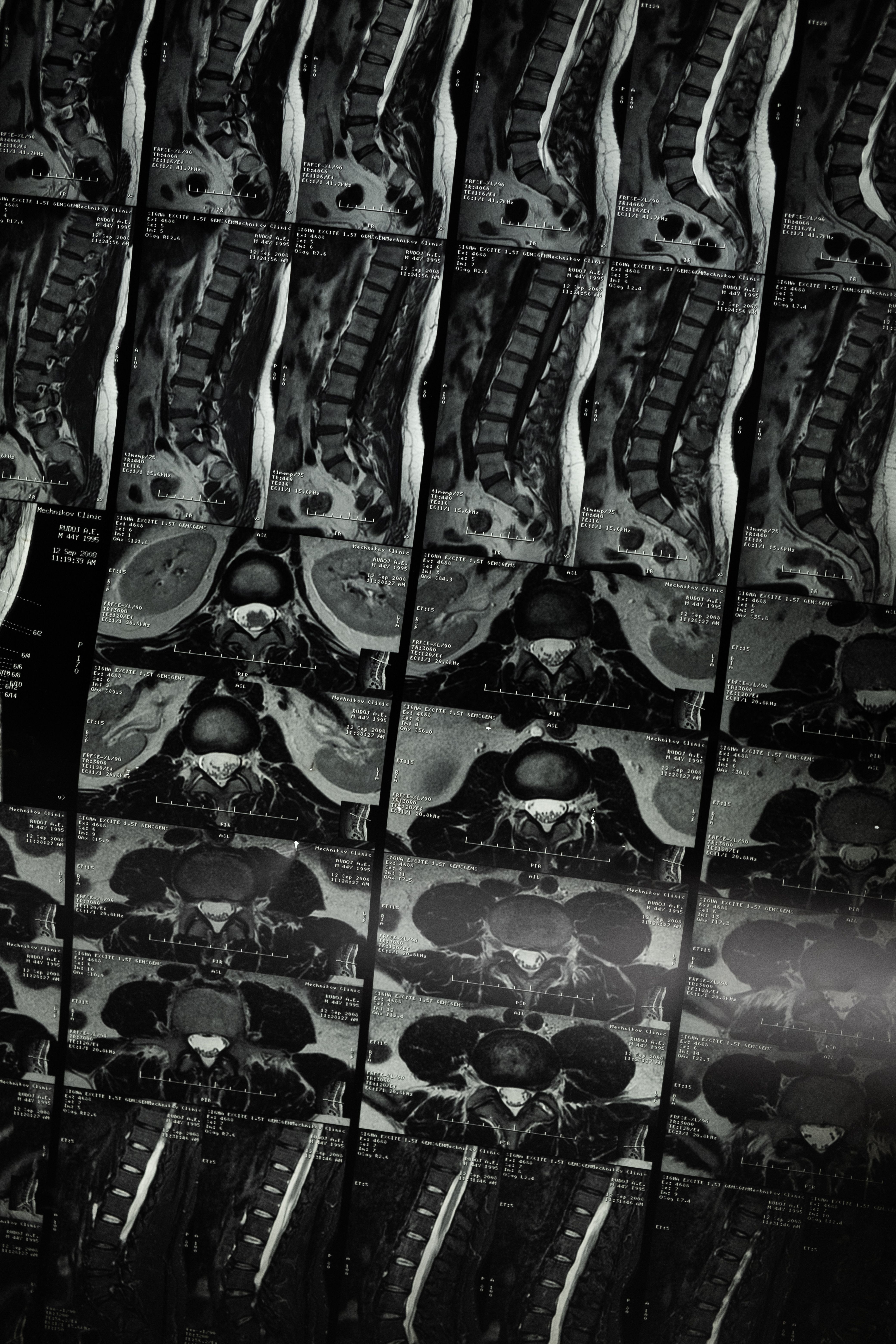
Magnetic resonance imaging (MRI)
Excellent for soft tissues
MRI delivers high soft-tissue contrast without ionising radiation
and is the modality of choice for soft-tissue, marrow, joint and
neurological questions. Multiplanar acquisition and a range of
contrast weightings make it central to musculoskeletal,
neurological, hepatobiliary and pelvic imaging, and it will
demonstrate pathology that CT resolves poorly or not at all. Its
constraints are physical rather than dosimetric. The static field
is permanently energised, so implant, device and foreign-body
screening is mandatory before entry to the controlled area, and
ferromagnetic projectile injury remains the principal safety
hazard. Acquisition times are long, motion degrades the study, and
a proportion of patients cannot tolerate the examination.
Gadolinium (an intravenous contrast agent) requires a documented
renal function assessment, and there are ongoing concerns about
tissue deposition in the brain. MRI capacity and cost continue to
limit access compared with CT. Its principal advantage is that it
carries no ionising radiation and therefore no IR(ME)R
justification requirement, but the request must still identify a
clinical question that MRI answers better than a faster or
lower-cost alternative.
Common indications
Joints and soft-tissue injury
Brain, spinal cord and peripheral nerve lesions
Liver, biliary and pelvic pathology
Bone marrow and musculoskeletal infection or tumour
Limitations
Implant, device and foreign-body safety
Long acquisition times, so motion degrades the study
Gadolinium contrast
Safety and practical notes
No ionising radiation, but the static magnetic field is always
on. Implant and device status must be checked before entry to
the controlled area. Gadolinium is avoided in significant renal
impairment unless clinically essential.
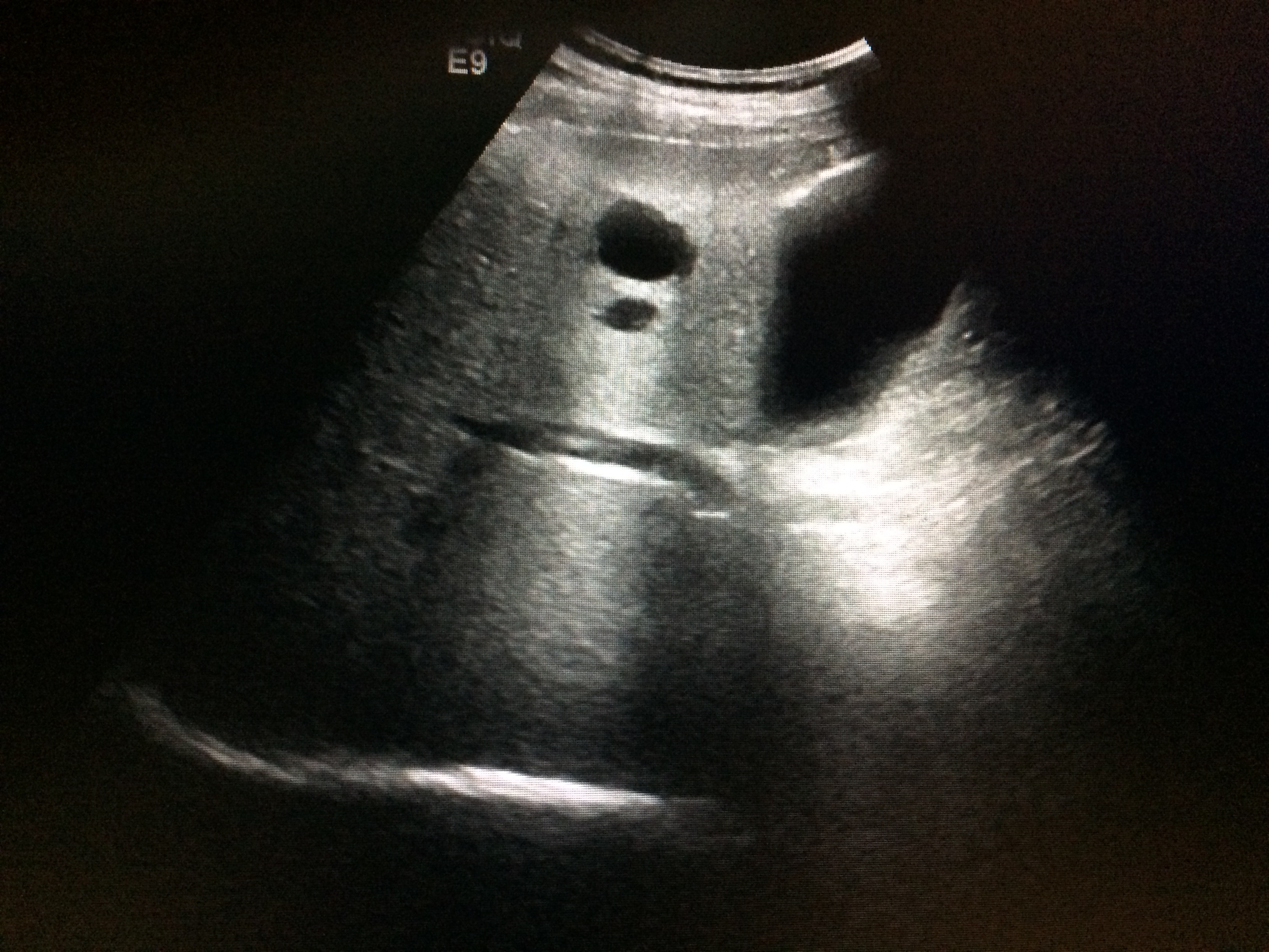
Ultrasound
Excellent for superficial structures and blood flow
Ultrasound provides real-time imaging without ionising radiation.
It is a dynamic examination rather than a set of static images, so
it can assess flow and superficial structures that projection and
cross-sectional imaging demonstrate poorly. It is widely used in
abdominal, obstetric, musculoskeletal and soft-tissue imaging, and
it is the guidance method for many interventional procedures. Its
principal weakness is operator dependence: image quality and
interpretation vary with the practitioner, and the examination is
the least reproducible of the modalities. Penetration is limited,
and bowel gas obscures deeper structures. Ultrasound uses no
ionising radiation, so no IR(ME)R justification requirement
arises.
Common indications
Abdominal and pelvic pain, including gallstones and renal
obstruction
Obstetric assessment and foetal wellbeing
Vascular stenosis, thrombosis and flow assessment
Image guidance for biopsy, drainage and vascular access
Limitations
Operator-dependent and the least reproducible modality
Limited penetration; bowel gas obscures deeper structures
No whole-body overview compared with CT or MRI
Safety and practical notes
No ionising radiation. Output parameters are kept as low as
reasonably practicable. Endocavitary and interventional probes
require appropriate decontamination between patients.
Figure 01
A radiographer checks a series of CT images on a monitor
No modality is better than the others: each has its own clinical use, with its own strengths and limitations.
§ 02 Dose & safety
Information on Dose, justification and statutory duties
Ionising radiation is a powerful diagnostic tool, but it carries a risk of harm. The statutory duties of justification and optimisation apply to every exposure, and the law requires that the dose be kept as low as reasonably practicable for the diagnostic purpose.
Figure 02
How the modalities compare on dose
Linear scale 0–20 mSv. MRI and ultrasound involve no ionising radiation.
UK average annual effective dose (all sources)
2.6 mSv
About half from radon; wide regional variation.
Average annual dose in Cornwall
7.8 mSv
Elevated radon; the highest regional average in the UK.
Public dose limit (man-made sources, excluding medical)
1 mSv/year
Statutory limit under the Basic Safety Standards.
Chest radiograph (for comparison)
≈0.02 mSv
A single projection; dose varies with technique.
Occupational dose limit (effective dose) i.e. radiation workers
20 mSv/year
Averaged over any calendar year; 100 mSv over five years under
IRR17.
Justification and optimisation keywords explained
Justification: The benefit of the exposure to the
patient must outweigh any potential harm. The referrer must provide
sufficient clinical information to allow the practitioner to justify
the examination and authorise the exposure.
Optimisation: Radiation doses are kept as low as
reasonably practicable (ALARP) while achieving the required diagnostic
purpose. The 2024 amendments to IR(ME)R strengthened duties to have
regard for diagnostic reference levels and to audit practice.
Diagnostic reference levels (DRLs): National values
derived from wide-scale surveys, typically set at the third quartile
of the dose distribution for standard practice. Local DRLs should
sit below national values, and departments consistently exceeding
them must review their practice and take corrective action.
Protocol choice: Ensuring the chosen study is capable
of answering the specific clinical question asked. A technically adequate
scan that fails to address the clinical query does not constitute
optimised care.
Who are the duty holders under IR(ME)R?
Employer: Establishes the organisational framework,
including written procedures, protocols, training, staff entitlements,
and compliance.
Referrer: Provides sufficient and accurate clinical
information to enable the justification of an exposure.
Practitioner: Justifies and authorises the individual
exposure by weighing clinical benefits against potential risks.
Operator: Carries out the practical aspects of the
procedure and ensures optimisation in practice.
The roles are distinct and may be held by different people for
the same exposure. A claim can attach to any one of them
independently.
What needs recording and reporting
Significant accidental or unintended exposures are reportable
incidents and must be investigated, with the findings recorded.
Occupational exposure is controlled through designated areas,
monitoring and dose limits. Under IRR17 a controlled area is
required where a person is likely to receive more than 6 mSv per
year, and a supervised area above 1 mSv per year.
Dose limits under IRR17 are 20 mSv per year (100 mSv over five
years), 150 mSv for the lens of the eye and 500 mSv for the
extremities.
Equipment must be commissioned, tested and withdrawn from use
when faulty.
QA records and dose records are evidence. They are routinely
sought years after the event, and their absence is itself a
finding.
Interpretation and reporting: the professional benchmark
The Royal College of Radiologists' Standards for interpretation and reporting of imaging investigations (third edition, October 2025) define what a defensible report looks like.
Report content
A report must answer the clinical question asked, describe the
findings, state the limitations of the study and give a clear
conclusion. A structured format is expected where one has been
agreed for that investigation. The conclusion should provide a
radiological diagnosis or a reasoned differential, acknowledge
limitations and make recommendations aligned with recognised
guidelines.
Named author and accountability
Every report must be attributable to a named reporting author,
including where reporting is remote or assisted by software. The
author's name, position and profession should be identifiable. The
content must not imply a level of scrutiny that has not in fact
been applied.
Remote and offsite reporting
Remote reporting is acceptable only where the reporter has the
images, the prior studies, the clinical information and the means
to communicate an urgent finding. The same expectations of
quality, turnaround and escalation apply as for onsite work. The
RCR standards for teleradiology set out the governance,
data-transfer and indemnity requirements.
AI-assisted and speech-recognition output
Where software generates or transcribes content, the named human
author remains responsible for its accuracy. Every automated
contribution must be reviewed and approved before the report is
released. The use of AI does not transfer accountability to the
software provider.
Turnaround and communication of critical findings
Providers must have procedures for turnaround times, communication
of unexpected or time-critical findings, imaging classification
for reporting priority, and acknowledgement of reports. A critical
finding must be communicated directly and the communication
documented, not left to the patient or referrer to discover.
Addenda and corrections
An addendum supplements the original report; it does not replace
it. It must be attributable, dated and linked to the original
study. Where a discrepancy or error is identified, the addendum
should be non-judgemental and should trigger the local discrepancy
and candour processes.
The standards apply to all who interpret and report imaging,
regardless of professional background.
A doctor reporting within a narrower scope of practice is held to
the same standard. Where imaged anatomy falls outside the
interpreter's expertise, radiologist review must be agreed in
advance. Reporting by sonographers and radiographers should occur
within consultant-radiologist-led teams, with supervision and audit
proportionate to the scope of practice.
Figure 04
Illustration for handling images section
§ 04 Handling images
Storage, transfer and viewing
Imaging disputes and clinical investigations frequently turn on digital provenance: what data was acquired, how metadata was handled, and who accessed the study. Rigorous image handling is an essential component of the clinical and legal record.
Anatomy of a DICOM study
A complete DICOM dataset comprises both pixel matrix data and an
immutable header containing metadata: patient and study
identifiers, acquisition parameters, exposure indices, display
presets, and slice geometry. This metadata enables searchability,
secure transfer, and anonymisation, while allowing subsequent
reviewers to distinguish between native acquisition features,
windowing choices, and actual pathology. Simply obscuring
identifiers on the visible image without stripping the DICOM
header leaves the file fully identifiable.
Secure transfer and anonymisation
When a study is transmitted outside the primary care pathway—such
as for specialist second opinions or medicolegal review—it must be
anonymised at source, transferred via secure encrypted channels,
and audit-logged. Royal College of Radiologists (RCR)
confidentiality standards distinguish between fully anonymised
data (stripped of all personal identifiers, dates of birth, and
facility tags) and pseudonymised data, which legally remains
personal data as re-linking keys exist. All transfers must comply
with institutional information governance protocols.
Second opinions and audit trails
The initiation of a second opinion, the credentials of the
reviewing clinician, and the formal advice rendered must be
documented within the clinical record. Where a secondary
interpretation diverges from the original report, formal
institutional discrepancy and escalation pathways must be invoked
to ensure patient safety and clear clinical accountability.
Cookies on this site
This site uses a single optional analytics cookie (Google Analytics 4) to
understand which reference pages are read. It is only set if you accept.
Strictly necessary storage — remembering this choice and keeping you signed
in — is always on. See the cookie policy
and privacy notice.